This decision was based on the comparison of mean FA and signal-to-noise ratios of an axial image at the C4–5 level for 4 healthy volunteers imaged on both scanners, which showed no significant difference in either parameter. The data from both the old and new MR scanner were included in the present analysis. The remaining 17 subjects were imaged using the following scanning protocol: 15 distinct directions, b-value of 600 sec/mm 2, TR/TE of 5000/98.2 msec, matrix size of 128 × 128, and FOV of 19 cm 2. The initial 6 subjects were recruited prior to May 2011 and were scanned on an older 1.5-T GE MR scanner using the following scanning parameters: 25 equidistant directions at a b-value of 500 sec/mm 2, TR/TE of 4500/80 msec, matrix size of 128 × 128, and FOV of 260 mm 2. Sagittal T2-weighted images of the cervical spine were acquired in all subjects by using a TR/TE of 4000/102 msec, matrix size of 384 × 224, and field of view (FOV) of 20 cm 2. 34 In the present analysis, we evaluate the relationship between FA determined from the high cervical cord (C1–2) and long-term functional outcome in patients with acute SCI.Īfter giving their consent, the patients underwent DTI of the cervical spine (C1–T1) on a 1.5-T MR scanner (Signa Excite GE Medical Systems) with a CTL spine coil. Previously, our group reported an association between FA value estimated at the high cervical cord (C1–2) during the acute phase of SCI and American Spinal Injury Association (ASIA) grades at admission, which showed the utility of DTI in early detection of structural changes in white matter tracts. 6, 11, 15, 27, 34, 35 One such DTI parameter, fractional anisotropy (FA), measuring the degree of diffusion anisotropy or restricted diffusion, is often correlated with injury severity and/or neurological outcomes. Studies in both acute and chronic SCI have demonstrated structural alterations remote from the site of injury in the form of changes to DTI indices. 2, 20, 23, 30 The restricted linear movement along the length of the spinal cord is due to barriers to diffusion such as axonal membranes and myelin sheath, which if interrupted manifest as alterations to the DTI parameters derived from the diffusion signal.

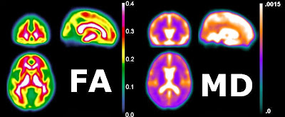
3, 13, 21, 22, 25ĭiffusion tensor imaging (DTI), a noninvasive imaging modality, uses the restricted diffusion of water molecules in neuronal tissue as the major contrast mechanism to provide quantitative information about microstructural integrity even within regions of the cord that appear normal on structural MRI.

The findings seen on anatomical MRI such as hemorrhage, lesion length, cord compression, and edema have been shown to have limited prognostic utility in predicting outcomes. Conventional MRI is most commonly used for the radiological evaluation of the injured cord. S pinal cord injury (SCI) is a devastating condition with high morbidity and mortality and is characterized by partial or complete loss of neurological function below the site of injury due to disruption of spinal cord integrity.


 0 kommentar(er)
